News | 01-03-2023
Three-dimensional visualization of lattice defects in single crystals
A combination of X-ray Diffraction Laminography (XDL) and Laser Scattered Light Tomography (LST) leads to the better understanding of the mobility of line defects.
Line defects or dislocations are a frequently observed phenomenon and represent a deviation from the strict atomic order of the crystal lattice. These defects influence the mechanical, electrical and optical properties of crystals as well as their growth behavior. For the development of high-quality crystals, it is therefore important to understand the origin and control the densities of dislocations. Although it depends on the specific application, dislocations have a detrimental effect on the crystals properties in most cases. [n1]
Once a dislocation is formed in the crystal, it cannot simply end, but will spread until it reaches the surface. Given sufficient temperature, however, dislocations can move due to internal stresses. This movement cannot take place in arbitrary directions but follows fundamental crystallographic rules based on different atomic mechanisms, denoted as glide or climb. Influencing this movement is thus one way to reduce the dislocation density in a crystal.
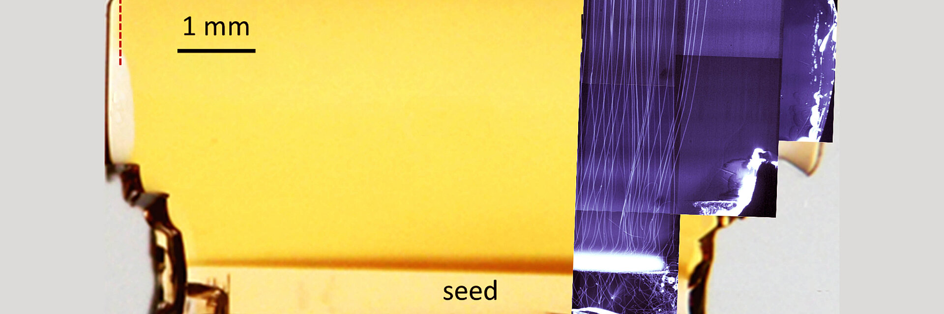
Three-dimensional (3D) imaging of the dislocation lines can provide valuable insight into the mechanisms of dislocation creation and their movement during growth. This way, scientist have indirect access to studying stress or impurity levels in the crystals and their shape as present during growth. This has been successfully demonstrated in a recent paper published by scientists from IKZ and Karlsruhe Institute of Technology [1]. Using 3D X-ray diffraction imaging they were able to recover the line shape and Burgers vectors of the dislocations in a aluminum nitride (AlN) single crystal obtained by seeded physical vaport transport growth at IKZ (T. Straubinger/Aluminium Nitride Crystal Growth) (Fig. 1).
The method, also called X-ray diffraction laminography (XDL), requires an intense, parallel as well as monochromatic X‑ray beam that is only available at synchrotron radiation sources (in this case PETRA-III, Beamline P23, Hamburg). Highly precise mechanics are furthermore needed, to rotate the crystal about a specific reciprocal lattice vector (Fig. 2[RDC2] ). Only this way, 3D information is accessible and a reconstruction of the illuminated volume is possible.
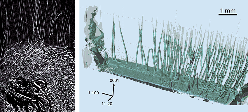
A more accessible technique to obtain similar information — laser scattering tomography (LST) — has recently been established at IKZ. While 3D XDL visualizes dislocations through their strain field, LST detects dislocations based on the fact that they are typically decorated by other atomic defects. These decorations locally enhance the scattering cross section of an incident laser beam, producing scattered light that can be detected by a camera. Lattice defects thus appear as a bright feature in an image. By scanning the crystal through the laser beam while continuously taking images, one then can reconstruct the 3D distribution as shown in Fig. 2. AlSince LST relies on the decoration of dislocations , there is a possibility that some may not be detected by this method. However, LST is much less demanding and allows the study of large volumes in comparatively short time. Combining LST and X-ray imaging thus is an efficient approach for 3D defect characterization in bulk single-crystals.
For further information, please contact Dr. Carsten Richter und Dr. Tobias Schulz
[1] T. Straubinger et al., Dislocation Climb in AlN Crystals Grown at Low-Temperature Gradients Revealed by 3D X-Ray Diffraction Imaging, Crystal Growth & Design 23, 1538 (2023).
http://doi.org/10.1021/acs.cgd.2c01131